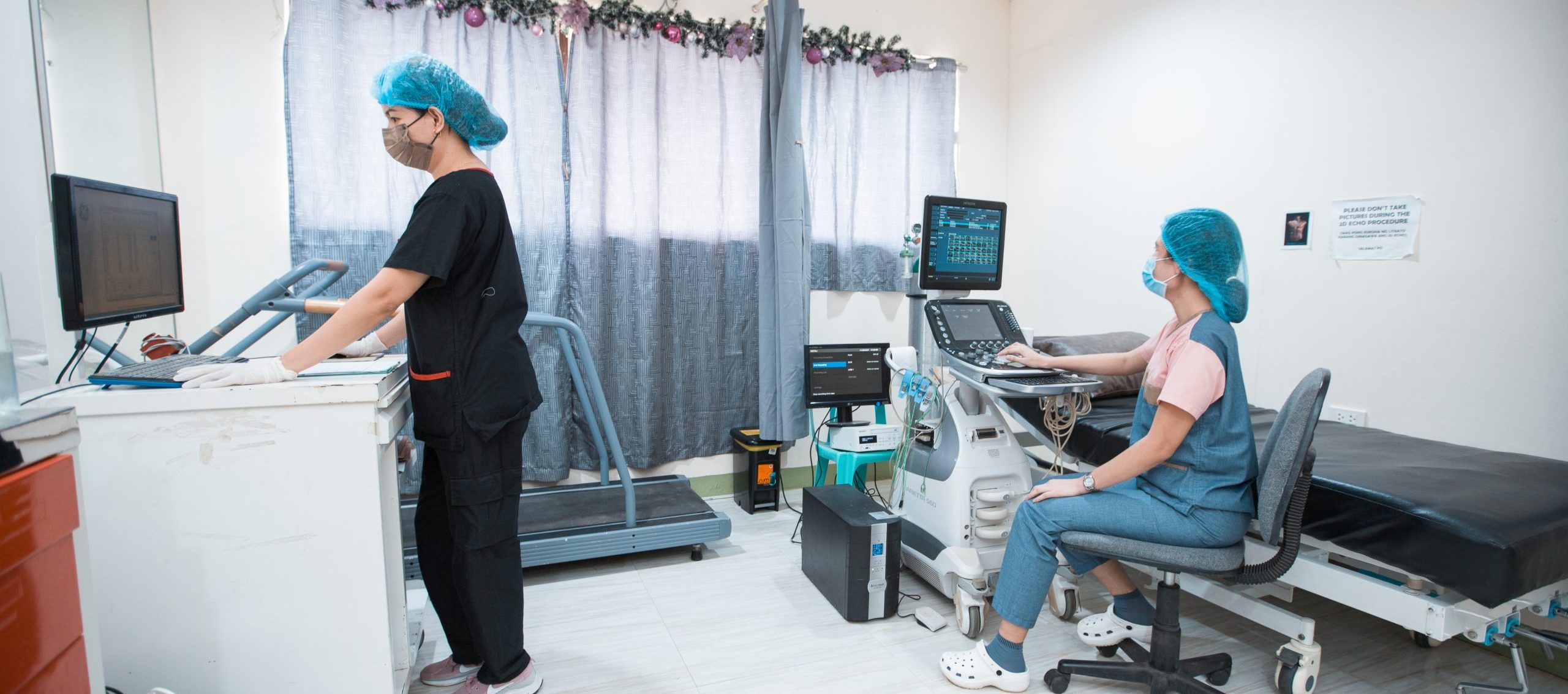
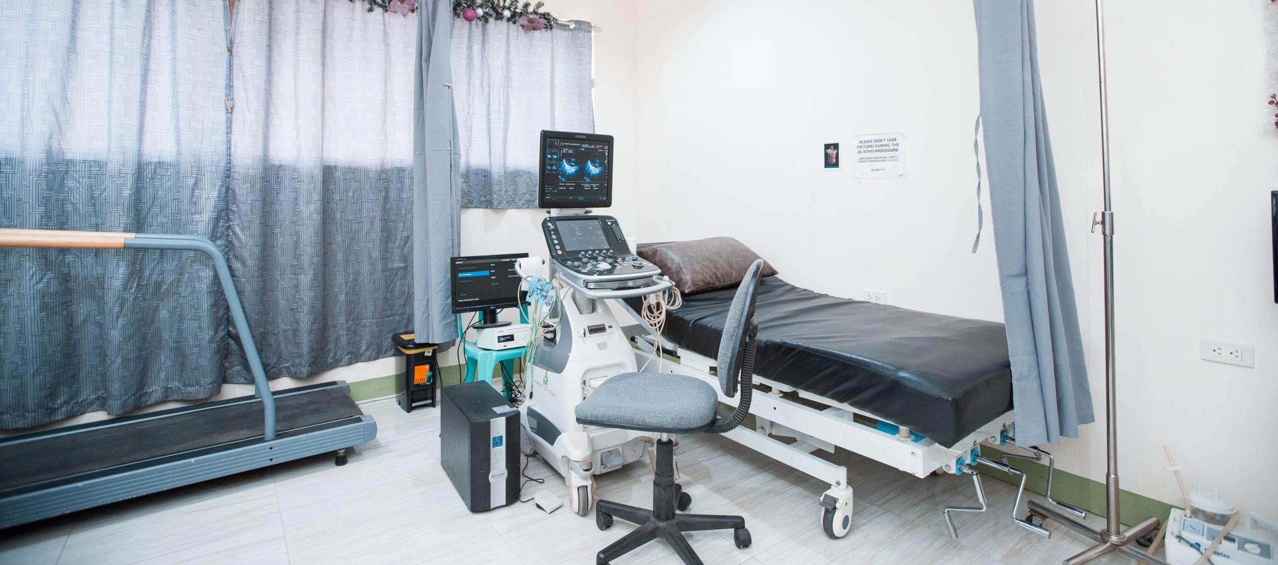
THE CARDIAC AND VASCULAR UNIT HAS FOUR CAPABILITIES:
1. 2D Echocardiography
2. Vascular Sonography
3. Treadmill Exercise Stress Test
4. 24hour Holter monitoring
MISSION:
The PMMGMPC Cardiac and Vascular unit is committed in providing competent patient care through accurate cardiac and vascular examination results rendered with professionalism and quality care.
VISION:
To provide competent patient care to the public by providing quality diagnostic cardiac and vascular examinations.
SERVICES OFFERED:
• 2D Echo with Color Flow Doppler
• Carotid Duplex Study
• Arterial duplex Study
• Venous Duplex Study
• Deep Vein Thrombosis Screen
• Abdominal Aorta Duplex scan
• Arteriovenous Fistula Mapping
• Arteriovenous fistula Evaluation
• Transcranial Doppler scan
• Treadmill Exercise Stress Test
• 24hour Holter Monitoring
• Newborn hearing screening test
Notes:
• Scheduling of procedures via phone call or text messaging.
• First come, First served.
• Operational hours: Monday to Friday 8 AM to 5 PM and STAT orders after 5 PM onwards, and on Saturday and Sundays
PROCEDURE DESCRIPTION:
A. Echocardiogram
• Utilizes ultrasound in the investigation of cardiac anatomy and function
• Safe, non-invasive technique
• Comprises of two-dimensional, real time imaging m-mode and spectral Doppler
1. 2D- Echocardiography plain
• Cardiac ultrasound in which the anatomy and patency of the valves are evaluated without the use of spectral Doppler.
• Thrombus, pericardial and plural effusion can be seen with this type of test.
2. 2D Echo with Color Flow Doppler
• Cardiac ultrasound with the use of color flow Doppler. It is a technique for recording the manner in which blood moves within the cardiovascular system.
• Doppler is a special part of the ultrasound examination that assess blood flow (direction and velocity).
B. Vascular Sonography
1. Carotid Duplex Study
A procedure that uses ultrasound to look for blood clots, plaque buildup, and other blood flow problems in the carotid arteries.
2. Arterial duplex Study
A procedure that assesses the arteries of the lower extremity.
3. Venous Duplex Study
A procedure that assesses the veins of the lower extremity consisting of the deep venous system, the superficial system, the dermal veins, the perforating veins, and the muscle tributaries.
4. Deep Vein Thrombosis Screen
A procedure in which it utilizes ultrasound to detect Deep vein thrombosis, or DVT, a blood clot that forms in a vein deep in the body. Blood clots occur when blood thickens and clumps together.
5. Abdominal Aorta
A procedure in which it utilizes ultrasound to detect Abdominal aorta abnormalities.
6. Transcranial Doppler scan (TCD) and Transcranial color doppler (TCCD)
Are types of Doppler ultrasonography that measure the velocity of blood flow through the brain’s blood vessels by measuring the echoes of ultrasound waves moving transcranially.
7. Arteriovenous fistula and graft Mapping
A procedure that assesses the arteries and veins for the creation of dialysis access. and also marking the possible site for the creation of the arteriovenous fistula and graft.
8. Arteriovenous fistula or graft Evaluation
When the fistula is created, the patency of the fistula or graft are evaluated and examined.
C. Treadmill Exercise Stress Test
A multi-phase exercise test for the purpose of finding indications of cardiovascular abnormalities.
A workload on the heart is gradually increased while the clinician observes the ECG, heart rate, blood pressure, as well as the signs and symptoms of the patient.
Phases:
-
-
- Resting Phase – the patient is asked to assume three different positions and vital signs are obtained.
- Exercise Phase – exercise proper wherein the patient walks and runs on the treadmill.
- Recovery Phase – the patient walks for a few minutes and sits on a chair until baseline vital signs are obtained.
-
D. 24-hour Holter monitoring
The Holter monitor is type of portable electrocardiogram (ECG). It records the electrical activity of the heart continuously over 24 hours.
POLICIES AND OBLIGATIONS:
1. Patients are scheduled in any agreed upon or available date. If there are no patients scheduled for the date, operations are managed via first come first served.
2. If the patient should have any preparation and doesn’t meet the exact requirements then he/she is scheduled again on another day.
3. The examination room should always be clean and prepared for the examination of the next patient.
4. Enough linen and patient’s gown is in stock to be prepared for the number of patients ahead.
5. Fresh linens should be changed as necessary.
6. There should be enough forms in stock for the day.
7. Ensures that enough tissue and ultrasound gel is in stock for the examination.
8. DVDs should be checked for capacity before placing it for recording.
9. The patient’s data should be written in the general entry logbook.
10. The reading physician should be informed immediately after the examination of the patient.
11. Results are obtained after 2 – 3 working days. However, if the situation demands results immediately, then the reading physician is informed to prioritize the result of the patient.
12. If there are outpatients whose cases demands immediate emergency interventions, then the reading physician is informed immediately.
13. Cleaning of the equipment in the examination room should be done every day or as necessary.
FOR PATIENTS WITH FINANCIAL ASSISTANCE FROM PCSO, DSWD, HMOS, ETC:
1. The request for the examination from the patient’s physician is checked to determine what type of procedure that needs to be done.
a. For patients asking assistance from PCSO, a price quotation is provided to the patient.
b. For patients with HMOS the approval for all the procedures should be first acquired from the HMOS office by the personnel from the admitting/information section.
2. If all the requirements are complete and verified by the billing or admitting/information personnel, then scheduling or exam proper can now be done.
PROCESSING OF OUTPATIENT INDIVIDUALS:
1. The patient shall present the request for the performance of the procedure from his/her attending physician.
2. The information clerk on duty shall encode the patient’s data.
3. Pay to the cashier for cash basis and if payment is thru HMO’s, PCSO etc.; Approval from the HMO, PCSO shall be acquired first before the scheduling/performance of the procedure.
4. For the results, the patient will be contacted via text or phone call when the results are ready for release.
PROCESSING OF INPATIENT REQUESTS:
1. The Nurse assistant/Nurse on duty hands over the cost center of the patient.
2. The examination of the stop charged patient must be paid in cash prior to the duty procedure.
3. Obtain necessary information from the patient’s chart.
4. Inform the patient of the procedure to be done. Explain the procedure, its purpose, duration and the necessary preparations. Obtain vital signs as necessary.
For patients ok to be transported to the examination room:
• Before transport to the examination room, the nurse station is informed.
• Relatives/Folks of the patient can accompany the patient during the examination.
• The patient is then transported back to his/her room and the nurse station is informed of the transport.


