RADIOLOGY DEPARTMENT
Radiography is an essential tool in the field of diagnosis. It is the initial diagnostic examination being requested to evaluate fractures, dislocations, bone infections (osteomyelitis) and bone tumors. With the advancement in technology, the PMMGMPC replaced the conventional film-based radiography with Digital Radiography. With the introduction of the digital camera, pictures can now be viewed within seconds, be manipulated or deleted, be easily multiplied and shared electronically. The tedious loading, processing and developing of films are also omitted. Exposure to environmentally damaging chemicals and expensive films are also eliminated. Aside from orthopedic use, digital radiography is also vital for proper evaluation of routine chest films and better evaluation of Trauma X-ray series in emergency cases. With this technology, PMMG-MPC Hospital embraced digital radiography as an additional service in the Department of Radiology and is now available for both inpatient and outpatient services.
Mission:
The PMMG-RADIOLOGY mission is to contribute whole heartedly and faithfully in the support of healthcare services thru giving good diagnosis without reservation and embracing the mission.
To serve and extend the utmost quality healthcare for human need.
To contribute to the support of the healthcare services being provided by the cooperative.
Vision:
To become a globally accepted diagnostic imaging institution.
To maintain a high – Quality radiological assessment to physician health care with fidelity and integrity to our clients view as a globally competitive clinical center.
SERVICES OFFERED:
GENERAL RADIOGRAPHY
Medical imaging is the technique and process of imaging the interior of a body for clinical analysis and medical intervention, as well as visual representation of the function of some organs or tissues. X-rays can be used to examine most areas of the body. They are mainly used to look at the bones and joints. In some cases, a substance called a contrast agent may be given before an X-ray is carried out. This can help show soft tissues more clearly on the X-ray.
CT scan
Computed Tomography, also referred to as a CT or CAT scan, is an advanced radiological imaging modality in which an x-ray beam rotates around the patient. It can visualize the internal portion of the organs and separate overlapping structures precisely, producing cross-sectional images of all parts of the body. It takes pictures that show very thin “slices” of your bones, muscles, organs and blood vessels so that healthcare providers can see your body in great detail.
Ultrasound
Diagnostic ultrasound, also called diagnostic medical sonography, is an imaging method that uses high-frequency sound waves to produce images of structures within your body. The images can provide valuable information for diagnosing and treating a variety of diseases and conditions.
ECG
An electrocardiogram is a simple test that can be used to check your heart’s rhythm and electrical activity. Sensors are attached to the skin are used to detect the electrical signals produced by your heart each time it beats.
MRI
An MRI (magnetic resonance imaging) scan is a test that creates clear images of the structures inside your body using a large magnet, radio waves and a computer. Healthcare providers use MRIs to evaluate, diagnose and monitor several different medical conditions. A medical imaging procedure that uses a magnetic field and radio waves to take pictures of your body’s interior. It is used to investigate or diagnose conditions that affect soft tissue such as tumors or brain disorders.
EQUIPMENT AND OTHER MODALITIES
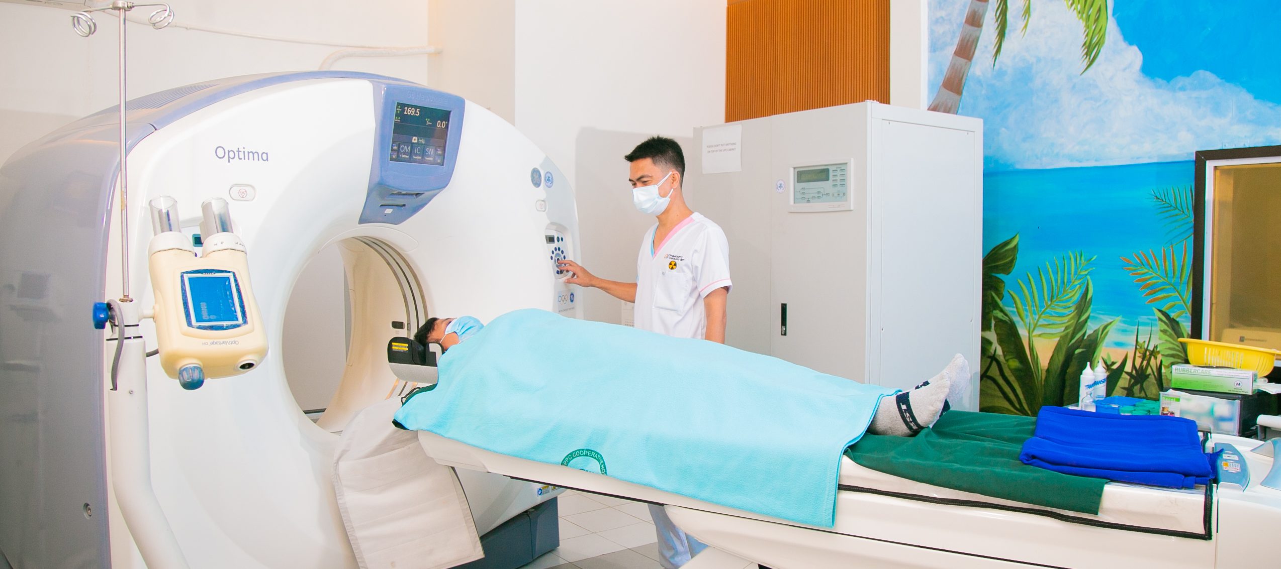
The GE Optima CT660 128 Slice CT scanner
The next evolution beyond the Bright speed and Discovery series, which continues on the themes of saving on space, dose, and cost. The GE Optima CT660 offers fast, high-quality image acquisition at optimized doses for highly competent, personalized care across a wide spectrum of procedures including cardiac angiography, neuro, abdomen, and much more. This GE Optima 660 128 Slice CT scanner for sale offers One-Touch Setup, allowing clinicians to personalize imaging, automatically apply preset advanced processing, volume-rendering, multi-planar reformats, and image sizing protocols.
- High Quality Images
- Fast Exam Time
- 500 lb. Table Weight Limit
- Seamless Clinical Workflow
- Ultra-Compact, Air-Cooled Helios Gantry
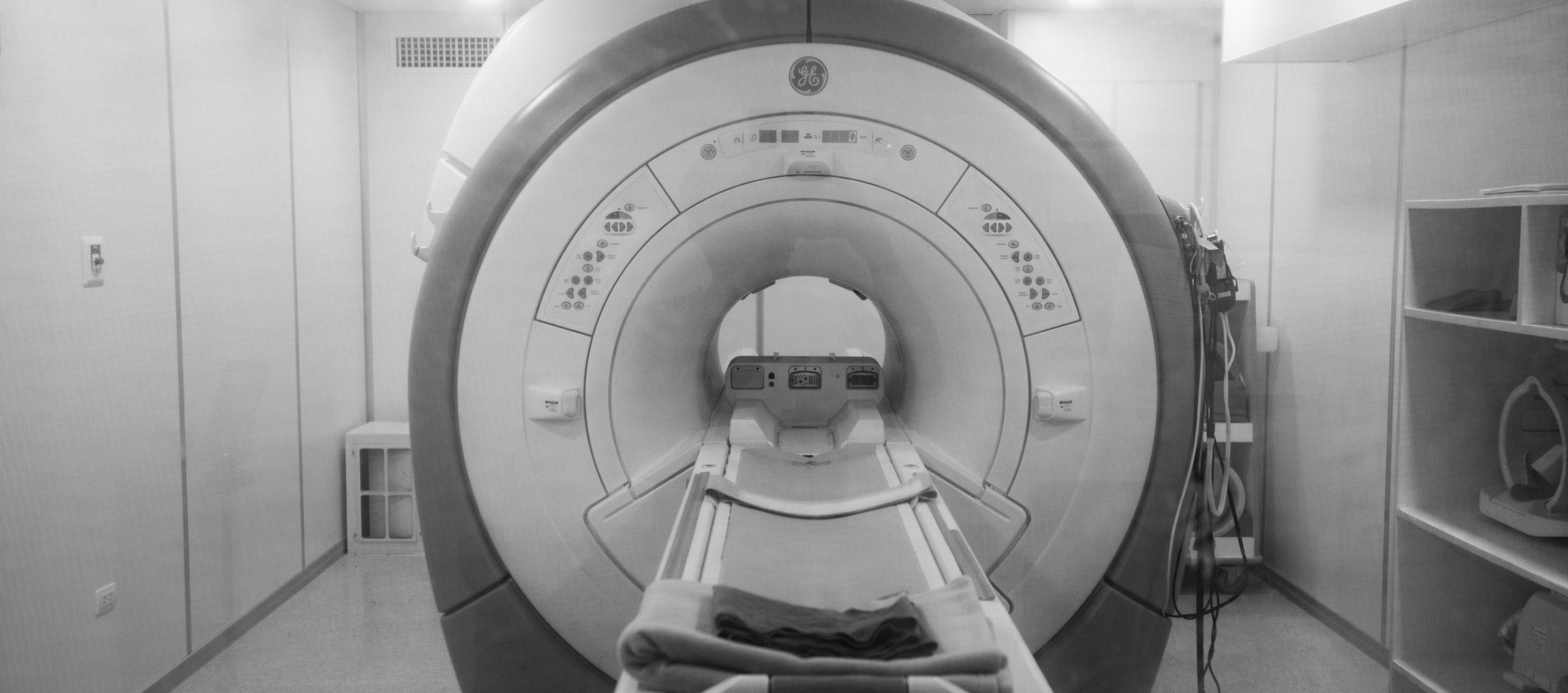
GE Signa HDxt 1.5 Tesla MRI
The GE Signa HDxt 1.5 Tesla MRI is geared completely for productivity.
The system empowers you more with consistent, high-quality imaging by overcoming fat sat failures, tissue characterization, and artifact reduction. The GE Signa HDxt 1.5T MRI also boasts reduced re-scanning/redundant exams due to patient or organ motion.
- Proven, Homogeneous 1.5T Magnet.
- 60 cm Bore
- 500 lb. Table Weight Limit
- HD Gradients Engineered for High-Fidelity
- HD Reconstruction Engineered for Real-Time, High-Performance Image Generation
- Advanced, High-Definition Applications
- High-Density Coils
The GE Signa HDxt 1.5T MRI is the upgraded, advanced version of the GE Signa HD, featuring an open design that provides outstanding lateral and medial access for patient comfort and safety.
MINDRAY RESONA 6 ULTRASOUND MACHINE
ZST+ powered premium image quality, Resona 6 provides essential clinical features with proven performance, intelligent total solution and intuitive gesture-based operation. All of these above have built up Resona 6 as the achievable premium solution to address challenges of clinical accuracy and efficient diagnosis in today’s demanding and overburden hospital environments. The channel data based ZST+ is an extraordinary innovation, representing an ultrasound evolution. Transforming ultrasound metrics from conventional beamforming to channel data-based processing; ZST+ is able to deliver multiple imaging advances: Advanced Acoustic Acquisition, Dynamic Pixel Focusing, Sound Speed Compensation, Enhanced Channel Data Processing and Total Recall Imaging.
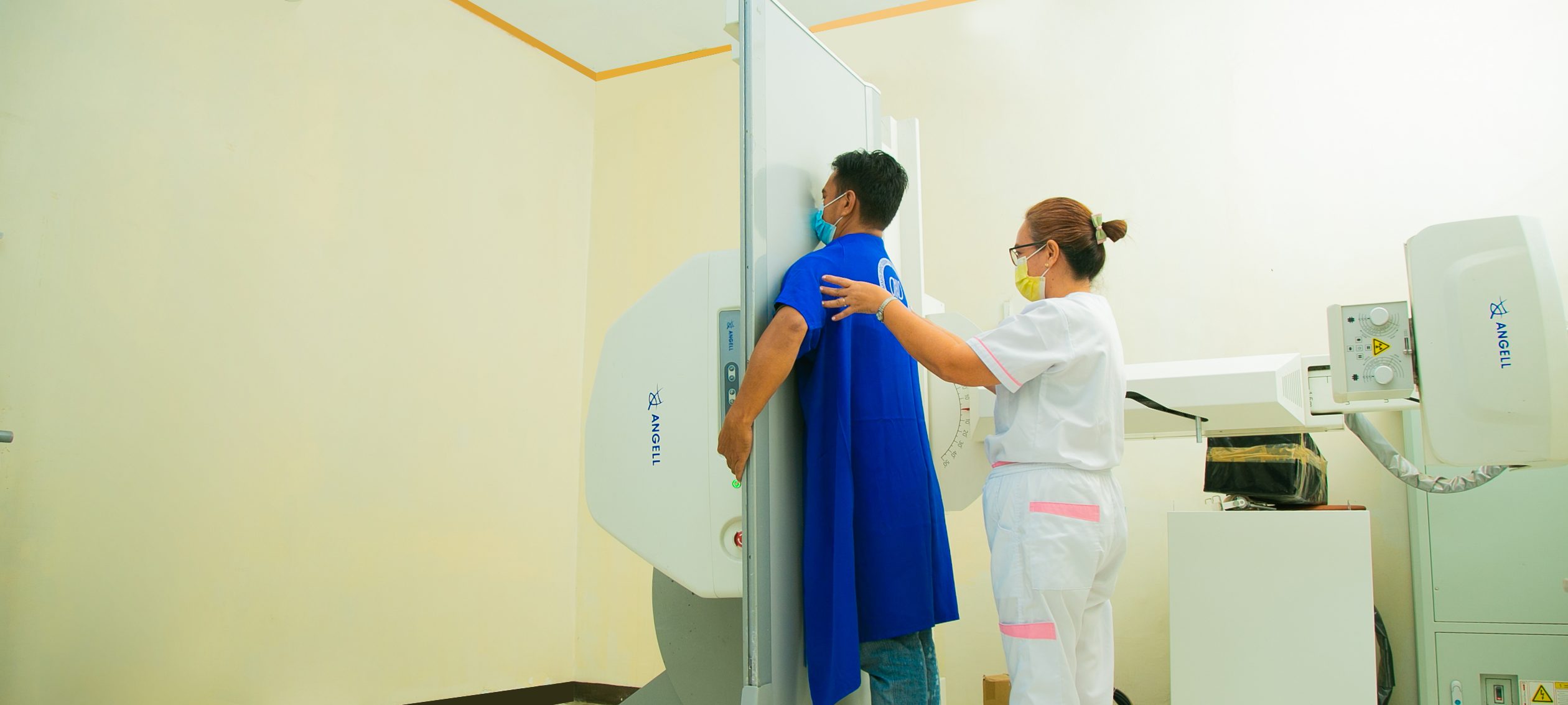
DYNAMIC DR X-RAY MACHINE
Digital X-ray radiographic technology has a wide range of applications in clinical image evaluation. From traditional X-ray radiographic technology to today’s digital X-ray radiography, the sharpness and accuracy of images have been greatly improved. In addition to digital X-ray radiography, dynamic DR radiographic technology has further broadened the thinking and practice of clinical application evaluation of X-ray radiography. Dynamic DR, through innovative technology fusion methods, enables X-ray radiography to break through the long-term imaging limitations of anatomical structures and enter motion functional imaging, providing important clinical value for imaging evaluation of diseases related to respiratory and joint systems.
OPESCOPE ACTENO C-ARM MACHINE
ACTENO incorporates functions to maintain high image quality and reduce X-ray exposure, even during long surgical procedures, such as Pulsed fluoroscopy, Multi Beam Hardening (MBH) filters, and Virtual Collimation. Furthermore, the area dose value is calculated and always displayed in real time on the touchscreen and monitor.
1M high-resolution CCD camera combined with our advanced imaging technologies delivers the required high image quality to you.
X-ray conditions can be easily set with a large Touchscreen on C-arm unit, where you can also select / change Simple-mode or Expert-mode. The screen can also show the fluoroscopic images as an optional feature.
Sports Medicine and Human Performance Center


