Microscopic Neurosurgery

MICROSCOPIC NEUROSURGERY
A First of its kind in Palawan
By Dr. Jesus Nigos, Neurosurgery
The operating microscope is a fixture of modern surgical facilities, and it is a critically important factor in the success of many of the most Complex and difficult surgical interventions used in medicine today. The rise of this key surgical tool reflects advances in understanding the principles of optics and vision that have occurred over centuries.
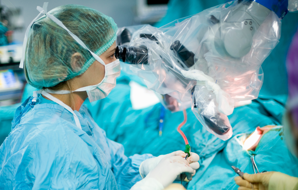
Always on the lookout for serving the need of the community and the province of Palawan, the PMMGMPC Hospital, in leading the way to medical excellence acquired a state-of-the-art surgical operating microscope, the acquisition of which pushes the boundaries of surgical care and the surgical experience to the next level. Microscopic Neurosurgery is now available in our institution, the first of its kind in Palawan.
Neurosurgery or Neurosurgical surgery is the medical specialty concerned with the prevention, diagnosis, surgical treatment, and rehabilitation of disorders which affect any portion of the nervous system including the brain, spinal cord, peripheral nerves, and extra-cranial cerebrovascular system. Our institution is fortunate to have a brain and spine surgeon who uses this advance in the field of microsurgery. He is no other than Dr. Jesus Nigos from Baguio General Hospital.
The Training Division Nursing Service Department, Palawan MMG Multipurpose Cooperative Hospital invited Dr. Jesus Nigos last March 10, 2017 to discuss about Microscopic Neurosurgery where he showed us the actual video footage of clipping of aneurysm perform in MMG’s state of the art operating Room. The lecture was conducted at the Medical Arts Building Conference Room of Palawan MMG Multipurpose Cooperative Hospital and Attended by almost 60 personnel.
A First of its kind in Palawan
A 62 y/o hypertensive female was brought to the ER after being found unconscious at their kitchen floor. She was preparing her family’s breakfast when she complained of a sudden severe headache different from some recurrent headaches she had in the past week. Her daughter claimed that she had an episode of seizure before being brought to the ER. A plain cranial CT scan showed a left sided subarachnoid hemorrhage. A CT cerebral angiogram shoed a left 12mm paraclinoidal aneurysm directed superiorly and laterally. A craniotomy with microscope assisted clipping of the aneurysm was then performed successfully. Microscopic surgery was introduced into the operating room by Dr. Carl Nylen, a Norwegian Otolaryngologist, in 1921. By 1942, Dr. Hans Hinselmann, a German Obstetrician, used a “Colposcope” in visualization of the inner female reproductive system. In 1953, Dr Hans Littman designed the first operating room microscope which was eventually used by Ophthalmologists, Plastic surgeons, and Vascular surgeons.
Dr. Theodore Kurze and Dr. Robert Rand’s Application of the microscope to neurosurgery in1957, radically changed the practice of neurologic surgery. It allowed mortality rates and complications to plummet. This tool had revolutionized concepts, allowed access to the deepest brain areas through efficient and excellent magnification combined with uncompromised lightning. Moreover, it expanded the boundaries of surgical techniques on minimally invasive neurosurgery. Improvement and perfection were reached and consequently mirrored in patient outcomes following surgical intervention. With the support of Dr. Alvin Timbancaya and the approval of the board of directors, the Leica Microsystems M525 F40 operating Microscope was acquired.
Finally, in January 5, 2017, the first microscope assisted brain tumor surgery (cerebellopontine angle acoustic achwanomma) was performed in Palawan MMG Multipurpose Cooperative Hospital. A first of its kind in Palawan. This was soon followed by a series of microscope assisted surgeries for hemorrhagic stroke cause by ruptured aneurysms (Clipping), for deep seated brain tumors (intraventricular Ependymoma) and other classic neurosurgical cases requiring detailed microscopic techniques. It has resulted in intense changes in the use and selection of instruments (micro instruments) and in the way operations were performed.
This Machine provides high quality optics and engineering for cranial, spine, multi-disciplinary, and other microsurgery disciplines (ENT, Ortho, Vascular, Plastic). These premium neurosurgical microscopic solutions feature brilliant illumination and magnification and has customizable options to offer the quality and reliability the surgeon needs. Its availability has greatly lessened the burden of families transferring the patients to other hospitals outside of Palawan.
Microneurosurgery, has improved the technical performance of many standard neurosurgical procedures, in both brain and spine, and has opened new, previously unattainable areas to the neurosurgeon. Operative results are hence improved by permitting faultless visual delineation of vascular and neural structures, deep seated areas in the brain are accessed with minimal brain retraction, smaller skin and cortical incisions, precise coagulation of bleeders and less damage to surrounding neural structures, more frequent preservation of compromised nerves in tumor cases. Likewise, small vessels and nerves can be anastomosed and sutured back, which has never been done without magnification. It has introduced a fresh era in clinical surgical education to the medical and nursing personnel by allowing observation and recording of minute surgical details not ordinarily visible to the naked eye.
